CHALLENGE
Restore an edentulous jaw with a subperiosteal implant using 3D technology and minimally-invasive surgery for improved results.
SOLUTION
- D2P® (DICOM to PRINT) software by 3D Systems
- Geomagic® Freeform® organic 3D design software by 3D Systems
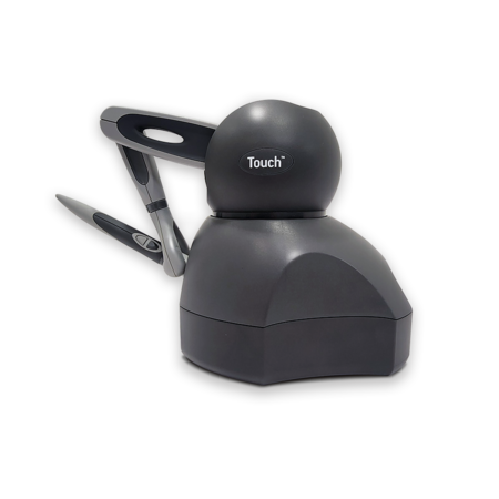
- 3D Systems Touch™ haptic device
- Plastic 3D printing (PLA)
- Metal 3D printing (medical-grade titanium alloy Ti6Al4V/Ti-64)
RESULTS
- Used surgical planning to facilitate the precise design of PSIs and screw position faster.
- Reduced repetitive drills during surgery.
- Improved accuracy.
- Eliminated multiple post-operative impressions and patient appointments, resulting in up to 60% time savings.
- Reduced rehabilitation time.
Graft3D Healthcare Solutions Pvt. Ltd. in Tamil Nadu, Chennai, India, is a reseller of 3D Systems software solutions for healthcare and also provides best-in-class design and metal 3D printing of patient-specific implants (PSIs). Graft3D has some of the country’s leading PSI design engineers and a team of medical experts including experienced surgeons. Graft3D specializes in creating individualized implants for complex surgical procedures using 3D solutions.
Rebuilding any anatomical defect is a difficult skill that involves surgeons manually shaping modeling, and placing bone cement, bone grafts, or titanium meshes. Surgeons can use PSIs to correct complex, symmetrical anatomical defects and tailor healthcare solutions for greater accuracy and shorter rehabilititaiton times. Dental work is a prime candidate for PSIs.
For example, a 50-year-old female patient was having difficulty chewing due to the loss of her posterior teeth. Subperiosteal implants are ideal for cases such as atrophic maxilla and mandible ridges. Using D2P software, Geomagic Freeform organic 3d design software, and 3D printing for the anatomical model and implants, Graft3D restored her edentulous jaw with a subperiosteal implant and minimally-invasive surgery while improving results. Surgical planning facilitated the precise design of the implant and screw position, which reduced repetitive drills during surgery.
Surgical Planning
Graft3D CEO V. Iraimudi, B.E, M.S. (by research), designed and analyzed the patient-specific implant. Dental surgeon Dr. C. Viha Preetha, BDS, processed the images and planned the surgery virtually with maxillofacial surgeon, Dr. B. Karthick, MDS.
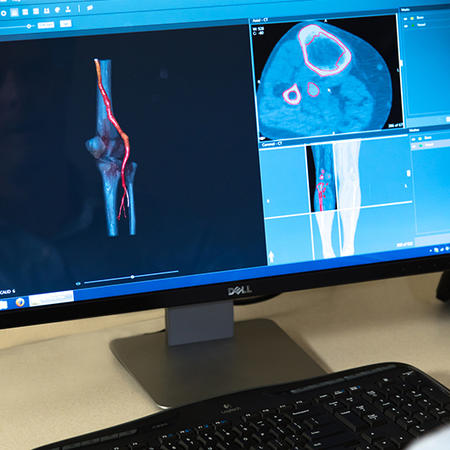
Conversion of DICOM to STL file in D2P software
Image Processing with D2P Software
Dr. Viha Preetha referred the patient for a CBCT (cone beam computed tomography) scan of both maxilla and mandible with less than 0.6 mm slicing thickness. She rendered the image in DICOM (digital imaging and communication in medicine) and then converted it into an STL file using D2P software from 3D Systems, which provides precise information including bone density and soft tissues and has a United States FDA 510(k) device clearance, European Economic Area (EEA) CE mark, and Israeli AMAR for medical use. Using D2P, she extracted the exact amount of bone exposed during CBCT radiation to perform an accurate and minimally invasive osteotomy.
Customized implants and anatomical model designed in Geomagic Freeform
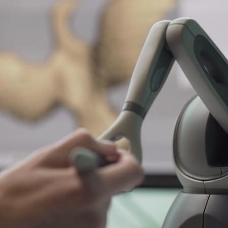
Surgical Planning and Designing with Geomagic Freeform Software
Next Mr. Iraimudi virtually planned and designed customized subperiosteal implants for every step of the procedure using Geomagic Freeform software and a Touch haptic device.
Mr. Iraimudi imported the converted STL file into Geomagic Freeform as an exact virtual replica of the patient’s jaw. He then used Geomagic Freeform and a Touch haptic device to design the implant framework accurately for location, length, depth, and angle using the interocclusal distance to position the abutment length and angulation for the prosthetic replacement.
By superimposing the pre-operative impression over the mandibular STL file in Geomagic Freeform, he designed a custom implant and saved time by assessing the procedure 50–60 percent faster, avoiding clinical obstacles, and eliminating multiple post-operative impressions and patient appointments.
3D-printed plastic anatomical model of patient’s jaw with customized metal implants
3D Printing Anatomical Model and Implants with Plastic and Metal AM
Next Mr. Iraimudi used the mandibular STL file to 3D print an anatomical model of the patient’s jaw and customized implants for the surgical procedure with a combination of plastic and metal additive manufacturing (AM) technologies.
He had the anatomical model of the patient’s jaw 3D printed on a plastic 3D printer with fused deposition modeling (FDM) technology using PLA. He had the customized implants 3D printed on a metal 3D printer using medical-grade titanium Ti6Al4V (Ti-64), which is compatible with human anatomy.
Modified ginwala incision and elevated buccal flap
Surgical Procedure
The prosthetic replacement operation was comprised of three steps that took place over a one-week period. The surgeon informed the patient of each step prior to surgery.
- Diagnosing and administering local anesthesia: Dr. Karthick diagnosed the patient with a posterior edentulous mandible and administered local anesthesia (inferior alveolar nerve block) to each side of the mandible.
- Performing incision and flap reflection: Dr. Karthick then placed an alveolar incision in the middle third of the premolar region ( modified ginwala incision) and elevated the buccal flap, relieving the underlying periosteum.
- Placing implants and screws and suturing: Finally, Dr. Karthick placed the customized subperiosteal implant on the edentulous ridge of each mandible side, placed the screws and adjusted the implants accordingly with minimal bone scraping, and closed the buccal flap with simple suturing.
Post-operative orthopantomogram
Result
Periodontal disease is the main cause of edentulous conditions. Bone loss occurs in infectious conditions, which can lead to standard dental implant failure. Clinicians can avoid such outcomes by customizing minimally-invasive, patient-specific subperiosteal implants through surgical planning. Using D2P and Geomagic Freeform software, clinicians can work interactively with patient-specific virtual models to simulate surgical procedures and improve the planning process. 3D-printed patient-specific implants are the bridge between a virtual platform and reality.